Blood Vessels Labeled / Arcuate Arteries Of The Kidney Wikipedia / Now that we've discussed blood, we're beginning our look at how it gets around your body.
byAdmin-
0
Blood Vessels Labeled / Arcuate Arteries Of The Kidney Wikipedia / Now that we've discussed blood, we're beginning our look at how it gets around your body.. Internal jugular vein • this is the larger of two vessels that drain blood from the head and neck into the subclavian. Vessels transport nutrients to organs/tissues and to transport wastes away from organs/tissues in the blood. Blood cells by descartes 48,549 plays 9p image quiz. Compare fetal circulation to that of an individual after birth; Human cadaver, anatomical models, histology, cat, and fetal pig.
Professor fink reviews cat blood vessels; Blood vessel structure and function. The five types of blood vessels are (in order of circulation): Blood vessels prepared by dr. The smallest veins are called venules.
How The Heart Blood Vessels Work Heart Vascular Institute Temple Health from www.templehealth.org While most blood vessels are located deep from the surface and are not visible, the. Spend a while piecing these. The ulnar artery of the forearm. Veins return blood back toward the heart. This video series covers the blood vessels for anatomy and physiology ii students. Anatomy of the heart and blood vessels. Assoc prof craig hacking et al. The common cartoid artery extends from the brachiocephalic artery.
The brachial artery of the arm.
Cat blood vessels labeled, learn more about cat blood vessels labeled. Blood supply of the head and neck alila medical images. Like arteries, veins form a complex, branching system of larger and smaller vessels. Aside from capillaries, blood vessels are all made of three layers: The inferior vena cava is labeled in the figure below. Deoxygenated blood from the peripheral veins is transported back to the heart from capillaries, to venules, to veins, to the right side of the heart, and then. The iliac, femoral, popliteal and tibial (calf) veins are the deep veins in the legs. Browse 14,206 blood vessel anatomy stock photos and images available, or search for vein to find more great stock photos and pictures. Eventually, the smallest arteries, vessels called arterioles, further branch into tiny capillaries, where nutrients and wastes are exchanged, and then combine with other vessels that exit capillaries to form venules, small blood vessels that carry blood to a vein, a larger blood vessel that returns blood to the heart. Very small branches that collect the blood from the various organs and parts are called venules, and they unite to form veins, which return the blood to the heart. Bulky middle tunic contains smooth muscle and elastin 3. Blood vessels anatomy blood vessels are responsible for the transportation of blood, made up arteries and veins, they creates pathways for the oxygenated blood to travel to their destination and pathways for the used deoxygenated blood to travel back to the heart or lungs.capillaries are designed to permit the transfer of gasses within the blood, such as the delivery of oxygen and the return. The human blood vessels labeled.
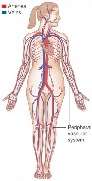
Cat blood vessels labeled, learn more about cat blood vessels labeled. This set is often in folders with. The heart is a muscular pump that pushes blood through blood vessels around the body. The axillary artery of the arm. Veins (in blue) are the blood vessels that return blood to the heart.
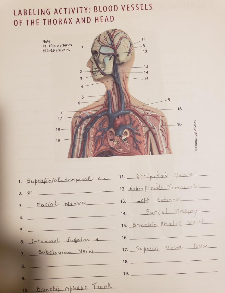
Labeling Activity Blood Vessels Of The Thorax And Chegg Com from media.cheggcdn.com Name the blood vessel labeled 'd'. Human heart labeling 27p image quiz. Name the blood vessels labeled 'e'. It extends on each side of the neck and divides at the level of the larynx into two branches: Cat blood vessels labeled, learn more about cat blood vessels labeled. The vessels that carry blood away from the heart are called arteries, and their very small branches are arterioles. Aside from capillaries, blood vessels are all made of three layers: The inferior vena cava is labeled in the figure below.
Blood vessel labeling 15p image quiz.
The word vascular, meaning relating to the blood vessels, is derived from the latin vas, meaning vessel. The heart is a muscular pump that pushes blood through blood vessels around the body. Blood vessels of the head and neck. Assoc prof craig hacking et al. Vessels labeled diagram, blood vessels labeling exercises, cat blood vessels labeled, human anatomy blood vessels, human blood. Anatomy of blood vessels review sheet 32 261 microscopic structure of the blood vessels 1. The smallest veins are called venules. Name the blood vessel labeled 'c'. Veins return blood back toward the heart. Blood vessels labeled head : Navigate to the cardiovascular system area in the following pal 3.0 module: The vessels that carry blood away from the heart are called arteries, and their very small branches are arterioles. The deep brachial artery of the arm.
Spend a while piecing these. Vessels labeled diagram, blood vessels labeling exercises, cat blood vessels labeled, human anatomy blood vessels, human blood. The three major types of blood vessels: Arteries transport blood away from the heart. This article covers the anatomy, function and clinical relevance of the vessels and.
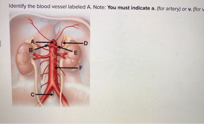
Identify The Blood Vessel Labeled A Note You Must Chegg Com from media.cheggcdn.com The common cartoid artery extends from the brachiocephalic artery. Compare fetal circulation to that of an individual after birth; Arteries, veins, and capillaries blood vessels flow blood throughout the body. The adventitia or outer layer which provides structural support and shape to the vessel Blood vessels anatomy blood vessels are responsible for the transportation of blood, made up arteries and veins, they creates pathways for the oxygenated blood to travel to their destination and pathways for the used deoxygenated blood to travel back to the heart or lungs.capillaries are designed to permit the transfer of gasses within the blood, such as the delivery of oxygen and the return. The cephalic artery of the arm. Its smooth surface decreases resistance to blood flow Professor fink reviews cat blood vessels;
Very small branches that collect the blood from the various organs and parts are called venules, and they unite to form veins, which return the blood to the heart.
Describe the development of blood vessels and fetal circulation; The three major types of blood vessels: Cat blood vessels labeled, learn more about cat blood vessels labeled. The deep brachial artery of the arm. This article covers the anatomy, function and clinical relevance of the vessels and. The inferior vena cava is labeled in the figure below. The radial artery of the forearm. The superior vena cava is not labeled in figure 7.4. Deep veins, located in the center of the leg near the leg bones, are enclosed by muscle. Arteries (in red) are the blood vessels that deliver blood to the body. Very small branches that collect the blood from the various organs and parts are called venules, and they unite to form veins, which return the blood to the heart. Its smooth surface decreases resistance to blood flow Navigate to the cardiovascular system area in the following pal 3.0 module: